The reviewed paper is Rapid-scanning forward-imaging miniature endoscope for real-time optical coherence tomography. One advantage of forward-viewing OCT endoscope over transverse-imaging counterpart is that it eliminates the need for mechanically actuating the entire endoscope assembly or a rotational fiber-optic coupler. Another possible merit is that the lateral-priority image acquisition sequence greatly reduces the depth-scanning speed.
The claimed “real-time” is achieved by the compact size and much faster lateral scanning of the device.
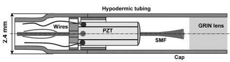
Fig.1 Schematic of the rapid-scanning forward-imaging miniature endoscope. (from the original paper)
As shown in Fig. 1, the proximal end of the GRIN lens was 8° angle polished to match the 8° angle-cleaved fiber tip surface. In addition, changing the distance between the fiber tip and the GRIN lens can fine-tune the working distance and the focused spot size. Multiplying the fiber tip scanning range by the magnification of the GRIN lens gives the lateral scanning range of the imaging beam on the focal plane.
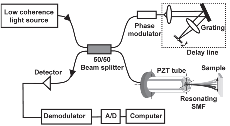
Fig. 2. Schematic of the fiber-optic endoscopic OCT system.(from the original paper)
Fig.2 shows the forward-viewing endoscopic OCT system. An electro-optic phase modulator is used in the reference arm to elevate the Doppler frequency shift to 1.5MHz, which results from a grating-based delay line. This frequency shift can be set to zero by centering the beam at the rotational axis of the tilting mirror. The delay line was set to compensate for the dispersion caused by the phase modulator crystal, as well as for slow depth scanning. For rapid lateral scanning, it is achieved by resonating the fiber cantilever with a tubular PZT actuator.
About the relation of confocal parameter b (twice the Rayleigh length) and waist radius w (half of the spot size), b=2\pi {w}_0^2/\lambda.
How to measure the confocal parameter?
Put a mirror in front of the GRIN lens, and measure the distance between the mirror at maximum intensity and 0.69*maximum intensity. Multiplying the distance by 2 gives the confocal parameter. At the position of Rayleigh length, the beam radius is √2w_0.