This old paper introduced two setups of forward-viewing instruments for optical coherence tomography (OCT).
In the beginning, the author mentioned one advantage of forward-viewing devices: permitting data to be collected before the device is introduced into the tissue. I assume it’s because of forward-viewing OCT’s characteristics.
Two setups were proposed: a hand-held probe, and a rigid laparoscope. Of course, I’m not going to list all the details of the devices, but only show some key points which are interesting or worth noting to me.
The sensitivity is measured as 110dB under the power of ~5mW on the specimen. Note that when we claim sensitivity, usually the power on the sample should be mentioned, otherwise the number of sensitivity can be confusing even misleading.
About the image formation of forward-viewing OCT, the laser beam was scanned on different transverse positions of sample, and the data set is a 2D crosss-sectional map of the optical back-scattering from the specimen. To realize full transverse image resolution, one must acquire longitudinal backscattering data (single A-line) with pixel spacing comparable to or less than the transverse spot size (to obtain oversampling of the image).
As shown in Fig. 1, changing the focal length of the second lens varies the magnification of the scan range in the object plane as well as the optical mode emitted by the fiber tip, therefore, changing the magnification scales both the focused spot size and the transverse scan length, making the ratio of the transverse pixel spacing to spot size constant. This is interesting.
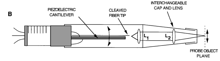
Fig.1. Probe telescope design for variable scan length and magnification.
About the coupling of single-mode fiber and GRIN lens, there is something worth noting. The faces of the 8°-angle-polished GRIN lens and the 8°-angle-cleaved fiber tip were angled to reduce internal back-reflections that would saturate the detector. The faces were oriented parallel to maximize coupling between the optical elements. This produced a 31-μm-diameter spot, which corresponds to a 1.2-mm confocal parameter, at a working distance of 3 mm from the GRIN lens (consider how to measure the confocal parameter, and calculate the spot size).
Reference:
[1] Forward-imaging instruments for optical coherence tomography